
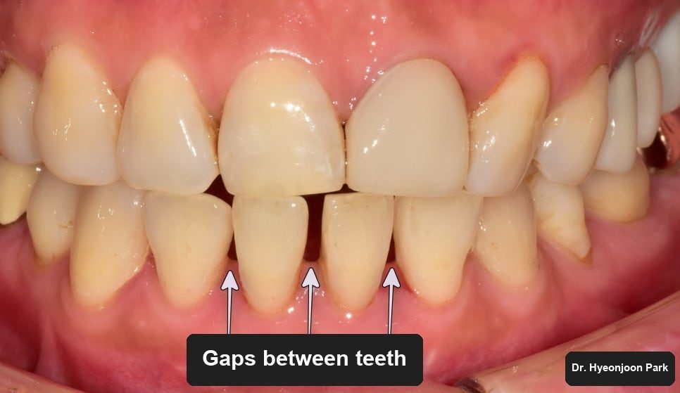
Gaps between lower front teeth are often closed with composite resin — it is quick, tooth-coloured and inexpensive. This case is what that repair can look like several years later, and what the X-ray found underneath it.
What the patient noticed
The complaint was appearance: the lower front teeth had become mottled and discoloured, with visible tartar between them.
Looking more closely, the shape of the teeth was not quite natural either. That was the clue. The spaces between these teeth — a diastema — had been closed some years earlier by building composite resin onto the sides of each tooth.
Why composite resin closes a gap only for a while
Composite resin is used constantly, and for good reason: it matches natural tooth colour closely and costs far less than laboratory-made work.
It has limits, and this case sits squarely inside them. Lower front teeth are narrow, so when the space to be closed is wide relative to the tooth, the added material makes the tooth look broad and flat rather than natural.
The second limit is time. Resin is not a permanent material. It needs maintenance, and where that does not happen it discolours at the edges, picks up staining and can eventually detach. That is what had happened here.
The black triangle
The old resin was removed and the teeth were cleaned. Straight away the mouth looked tidier — and straight away the original problem was visible again.
Between the teeth, at the gum line, sat dark triangular spaces. These are called black triangles. The gum tissue is supposed to fill that area; where the gum has receded, it no longer does, and the gap shows as a shadow.
This is the point at which building the space up with resin again would repeat the same outcome. Where black triangles are wide, prosthetic work — veneers, crowns or a bridge — gives a more stable and more natural result.
What did the X-ray find under the resin?
Before any of that, imaging was taken. That decision mattered.
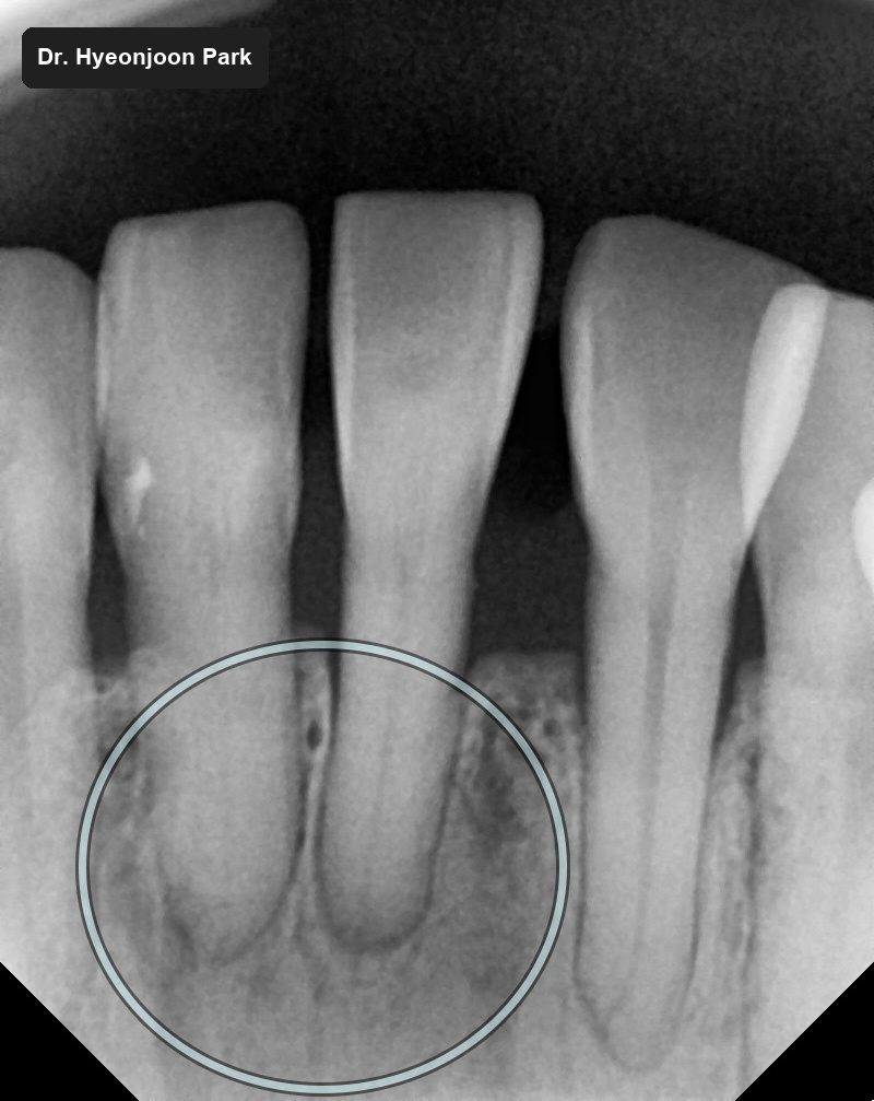
Gum recession this pronounced, in a mouth where cleaning had clearly been difficult for a long time, usually means there is more going on than can be seen. The X-ray confirmed it: a large area of infection around the root, with substantial loss of the surrounding alveolar bone.
That degree of bone loss means the tooth would already have felt loose. Caught early, it could very likely have been saved. Given the size of the lesion and the state of the bone at this point, it could not be.
This is the part worth taking from the case. Nothing about the outside of these teeth said “root infection”. The complaint was that they looked untidy.
Why remove a failing tooth promptly?
Where a tooth cannot be preserved, removing it promptly protects the teeth on either side, which are still healthy and are the next thing at risk.
An implant is the option most people expect, and it is not the only one. Where the neighbouring teeth are sound, a bridge uses them as supports and spans the gap with a single fixed piece of work.
That was the route taken here, and it had a second advantage in this particular mouth: because the bridge is made as one unit, the spacing across the front teeth could be designed into it. The diastema that had been patched with resin was resolved as part of the restoration rather than treated separately.
Temporary teeth and shade matching
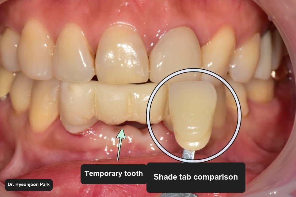
A bridge is made in a laboratory, which takes time, and nobody can go without front teeth in the meantime. A temporary bridge is fitted for the interval. It is less refined than the final work — it is not meant to be otherwise — but it restores appearance and function while the permanent one is made.
Before that, the colour is chosen. Shade taking means holding graded shade samples against the neighbouring teeth in the mouth and selecting the one that blends. That reading, together with how the lower teeth meet the upper ones, is what the technician works from.
What did the finished bridge look like?
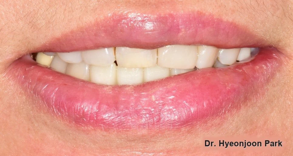
The finished bridge was fitted, the bite was adjusted, and the case was completed.
The spacing was resolved and the tooth shapes read as natural rather than built-up, and the lower teeth followed the lip line as they should.
How long does a front bridge take?
Examination on 14 February 2024, completion on 27 February 2024 — about two weeks, including the laboratory time for the bridge.
How do I make the bridge last?
Aftercare decides how long this lasts. Not only brushing: a restored area needs periodic professional examination, because most of what goes wrong underneath a restoration cannot be seen or felt from outside. That is exactly how this tooth was lost in the first place.
Frequently asked questions
What is a black triangle between teeth?
It is the dark triangular gap that appears at the gum line between two teeth when the gum tissue that should fill that space has receded. It is a gum-level problem rather than a tooth-level one, which is why simply adding material to the teeth often does not resolve it convincingly.
How long does composite resin last on front teeth?
There is no fixed figure — it depends on how large the repair is, where it sits and how it is maintained. What can be said is that it is not permanent: it discolours at the margins over time and can detach, and it is less predictable on narrow lower front teeth where a wide space has to be closed.
Can a tooth with root infection be saved?
Often yes, if it is found early. What decides it is how much of the surrounding bone has been lost. Where the supporting bone is largely gone and the tooth is mobile, preservation is no longer realistic and removal protects the neighbouring teeth.
Bridge or implant — which is better after an extraction?
Neither is better in the abstract. A bridge uses the adjacent teeth as supports and is completed in weeks; an implant stands independently but takes months while it integrates with bone. The state of the neighbouring teeth, the bone and your own schedule all feed into the decision, which is why it is made after the examination rather than before.
Will I be without front teeth while the bridge is made?
No. A temporary bridge is fitted for the interval between preparation and fitting the final one, so appearance and eating are covered throughout.
Read more: crowns and veneers · cavities and fillings · dental implants