
Anything covered by a restoration is, by definition, hidden. This case is what a gold bridge had been concealing — and how the extent of it was measured before anything was touched.
Why is a problem under a bridge hard to find?
Some problems in the mouth can be seen. Anything underneath a crown or bridge cannot.
X-rays are the first way in. Occasionally even those do not settle it, and the only remaining option is to remove the restoration and look — which is worth knowing before treatment starts, because it means the plan can change partway.
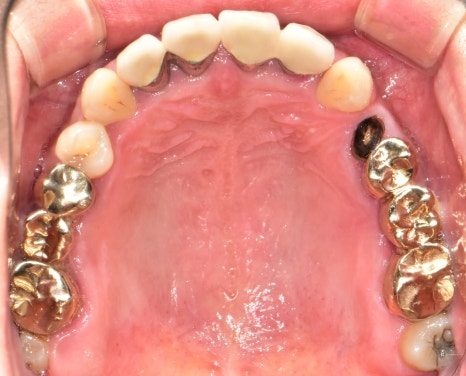
Here the visible finding was an old gold bridge spanning the back teeth. The complaint was gum pain, which in this context usually means periodontal disease.
Why does bone loss narrow the options?
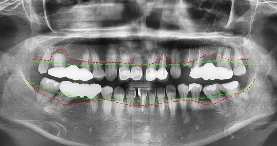
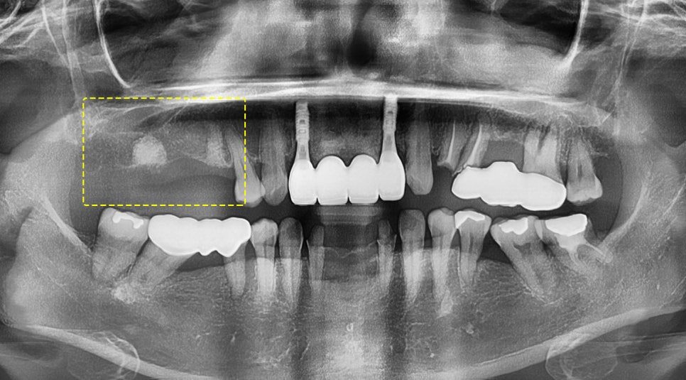
On the panoramic image the bone level can be assessed by comparison: where the alveolar bone should sit, against where it actually does.
The two lines did not run parallel. The bone had dropped and receded, unevenly, along the run of the bridge.
That measurement is what drives the plan, because severe bone loss can rule out the treatment a patient would otherwise have chosen. Options narrow as bone disappears, and they do not widen again.
Upper right: extraction and immediate placement
On the upper right the periodontal condition was advanced enough that the teeth could be extracted and implants placed at the same appointment.
That sounds counterintuitive — worse condition, faster treatment — but immediate placement depends on the state of the socket and the surrounding bone rather than on how healthy the tooth was.
The loose, weakened molars were removed together with the wisdom tooth behind them in a single procedure. Some grafting was carried out around the sites while avoiding the maxillary sinus above, and the fixtures were placed.
One point specific to the upper jaw: the bone there is softer than in the lower, so initial stability is harder to achieve. Where the fixture is positioned to get that grip matters more than it does below.
Why was the lower side placed with a guide?
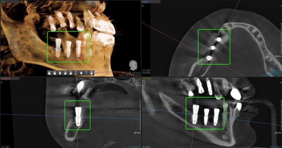
On the lower right, mobility was expected to follow the same course, so that side was treated with digital guided placement.
The reason is the inferior alveolar nerve running through the lower jaw. The fixture has to stay clear of it, and how much clearance exists is not something to judge by eye during surgery.
Planning on the scan data establishes the distance to the nerve, the density and thickness of the bone, and from those the placement position — and it settles the fixture diameter and length in advance. Deciding those before the surgery reduces error and shortens the time the site is open.
The post-operative images confirmed the fixtures sitting centrally within the bone, with graft material evenly distributed where it had been placed.
What happens once the implants have healed?
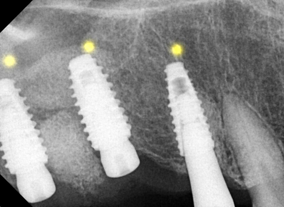
After the integration period, bone had filled in around the fixtures — visible on X-ray as increased radiopacity around them, which is the sign that the site has consolidated.
With the fixtures stable, zirconia crowns were fitted, chosen for durability under molar chewing loads.
Treatment ran from 7 May 2024 to 6 November 2024, about six months.
What happens if I put off treatment?
From the point at which teeth become mobile and painful, the alveolar bone continues to resorb. It does not hold at that level while a decision is made.
Every month of that makes the eventual treatment more complex: more grafting, more stages, and a narrower set of options to choose from. The difference between treating this at first symptoms and treating it a year later is not the same treatment done later — it is a different, larger treatment.
Frequently asked questions
How can I tell if I have bone loss around my teeth?
Not by looking. It is assessed on X-ray by comparing where the bone level should be against where it is. Loose teeth and gum pain are late signs; by the time they appear, the loss is usually well established.
Can an implant be placed on the same day as the extraction?
Sometimes. It depends on the condition of the socket and the surrounding bone, not on how bad the tooth was. Where it is possible it saves months; where it is not, placing early risks the implant.
Is the upper jaw harder to implant than the lower?
Different rather than harder. Upper bone is softer, so getting initial stability takes more care over positioning, and the maxillary sinus limits available height. The lower jaw is denser but carries the inferior alveolar nerve, which sets its own constraints.
Why do I need a new X-ray if I already have a bridge?
Because the bridge hides everything underneath it. The X-ray is the only way to assess the bone and the supporting teeth without removing the restoration — and occasionally it still has to come off before the picture is complete.
How long does this kind of treatment take?
This case ran about six months across both the upper and lower right. Most of that is integration time rather than appointments, so it can partly be spent at home if you are travelling for treatment.
Read more: dental implants · digital implants · bone grafting and sinus lift