
Two people can be told very different things about the same wisdom tooth, and the reason is usually what was imaged before the decision. This is the examination sequence, and what each stage is actually looking for.
The tooth in question
The wisdom tooth is the rearmost molar — clinically the third molar.
Kept clean, it can stay and function like any other tooth. The problem is that most of them cannot be kept clean. A toothbrush struggles to reach that far back, and where the tooth is angled or half-buried there are surfaces a brush physically cannot touch. Decay follows, and by the time it is noticed the removal decision has usually made itself.
What does the first examination check?
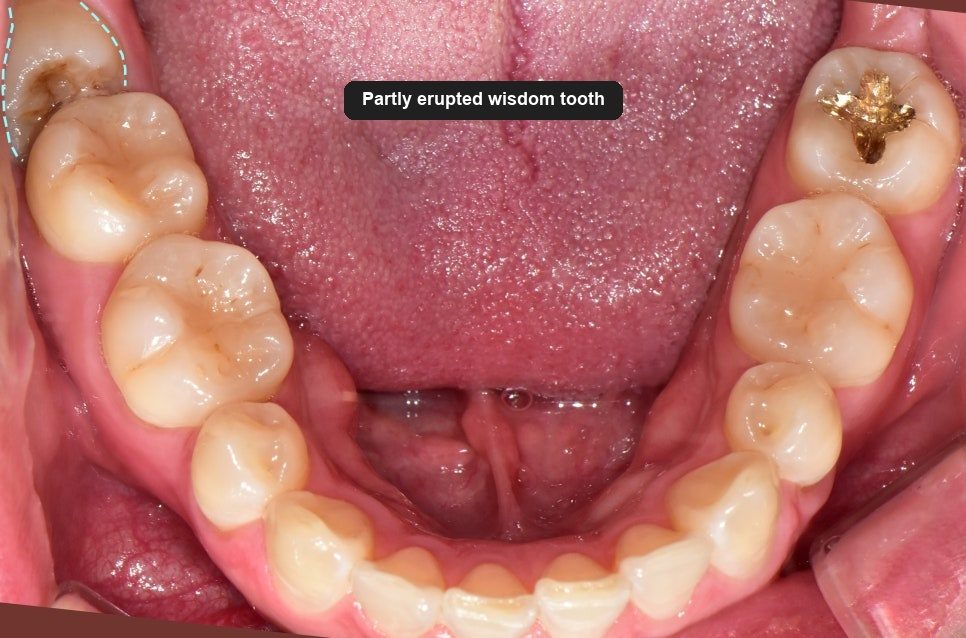
The first thing done is to look. How far the tooth has erupted, at what angle, whether decay has already started, and the state of the gum around it.
This is the stage that decides what imaging is needed, so it is not a formality.
What does the panoramic X-ray show?
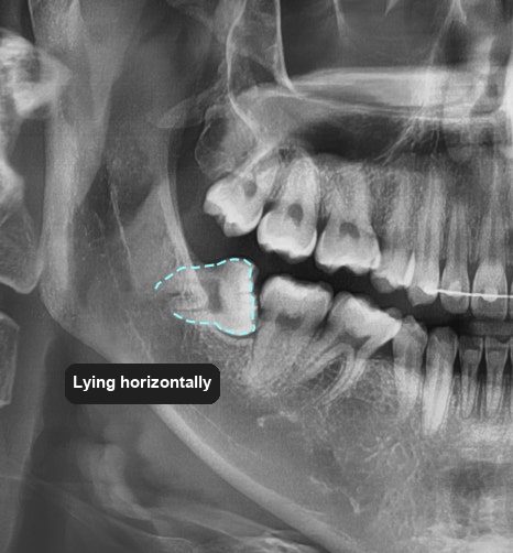
A panoramic film shows the whole jaw in one image, including the parts of the tooth that are still under the gum.
What matters on that image depends on where the tooth is. For an upper wisdom tooth, the relationship with the maxillary sinus. For a lower one, the inferior alveolar nerve canal — the channel carrying the nerve that supplies sensation to the lower lip and chin. If that canal runs close to the roots, removal has to be planned around it, because injury there can leave altered sensation.
What does a 3D CT show that an X-ray cannot?
If the roots and the nerve canal appear to overlap on the panoramic film, a 3D CT scan is added.
The word “overlap” is doing a lot of work there. A flat image cannot distinguish between roots that genuinely sit against the nerve and roots that merely pass in front of or behind it. Those are very different surgeries. The CT resolves which one it is.
The scan is added when the flat image raises the question — not as a matter of routine for every wisdom tooth.
How did the imaging change the plan?
The third molar here was partly erupted: some of the crown was through, and roughly half of it remained covered by gum. The panoramic film showed it lying at an angle rather than upright.
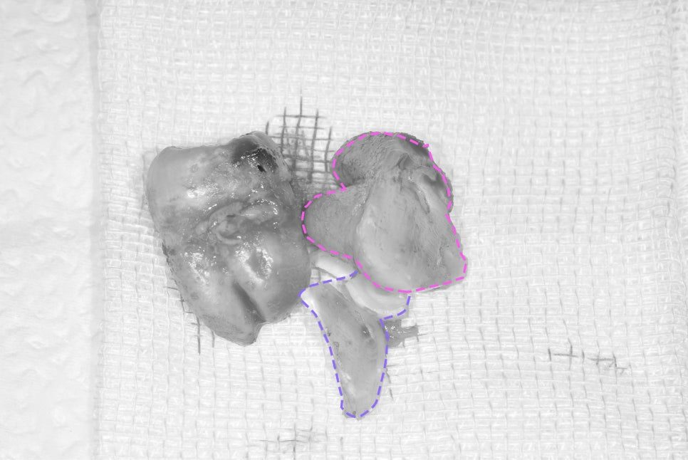
A partly buried tooth in that position has very little room to come out along its own axis. So the plan was to section the tooth — separate the crown from the roots and remove them as separate pieces, each through the space available, rather than forcing the whole tooth out in one.
The angled position also explains the decay risk directly. Plaque and tartar build up where the brush cannot reach, and food packs into the gap between the wisdom tooth and the molar in front, which inflames the gum around both.
What the plan avoided
An impacted wisdom tooth commonly requires the gum to be incised and laid back to reach it.
Here it did not. The tooth was not deeply impacted, so it was possible to remove it by sectioning alone, without a gum incision. It came out in two pieces.
That is the practical payoff of imaging first. Less soft tissue disturbed means less swelling and a shorter recovery — and whether it is possible is a finding, not a preference.
After the extraction
The gum was sutured closed. A dressing appointment follows the next day, to check for bleeding and assess swelling while it is still early enough to act on.
Sutures come out about a week later. If nothing unusual has appeared by then, the next check is a routine one at around three months.
Healing speed depends on the surgery, and it also depends on oral hygiene during the healing period. That part is not in the clinic’s hands.
The short version
The sequence looks elaborate for one tooth. It is what allows the removal itself to be quick and precise — and a quicker, more precise extraction is what produces less pain and less swelling afterwards. The examination is not the delay before the treatment; it is most of what determines how the treatment goes.
Frequently asked questions
Do I need a CT scan to have a wisdom tooth out?
Not always. It is added when the panoramic X-ray shows the roots overlapping the inferior alveolar nerve canal, because a flat image cannot tell whether they truly touch. Where there is clear separation, the panoramic film is enough.
Will my gum have to be cut?
It depends on how deeply the tooth is buried. Where it is only partly covered, it can often be sectioned and removed without an incision, which means less swelling. Deeply impacted teeth usually do require the gum to be opened.
Why is the tooth cut into pieces?
Because an angled or partly buried tooth has no clear path out as a single unit. Separating the crown from the roots lets each piece come out through the space that exists, rather than removing bone to create space for the whole tooth.
Can I keep my wisdom teeth?
Yes, if they have erupted upright, meet the opposing teeth properly and can be cleaned. The reasons to remove one are decay, damage to the molar in front, recurrent gum inflammation, or a position that makes any of those inevitable.
How many appointments does it take?
Typically the extraction, a dressing check the following day, and suture removal about a week later. If you are travelling, that spread is worth building into your dates.
Read more: cavities and fillings · emergency dental care · planning dental treatment in Korea