

You come in about the colour of one front tooth and leave with a plan involving a root canal and a crown. From the chair, that can sound like the treatment was talked upwards. Usually it was not. In a front tooth, colour change is a late sign of decay, and by the time it shows, the decay behind it has often already reached the nerve.
Why does a decayed front tooth change colour?
Two things darken a tooth from within, and decay produces both.
The first is the decayed tissue itself. Enamel on an upper front tooth is thin and partly translucent, so decayed dentine underneath shows through it as a grey or brownish shadow — which is why patients describe the tooth as looking bruised. The mark does not brush off and does not respond to whitening, because it sits under the enamel rather than on it.
The second is the pulp, the nerve and blood supply inside the tooth. Where decay reaches it the tissue inflames and then dies, and the tooth darkens from the inside outwards. Here the discolouration was pronounced enough that deep decay was suspected before any imaging.
Why does decay reach the nerve sooner in a front tooth than in a molar?
Because there is less tooth between the outside and the nerve. An upper front tooth is small, with a single canal down the root that is wide in proportion to the tooth around it. A molar carries far more bulk of enamel and dentine over its pulp, and its canals are narrower and usually three or four in number.
The consequence is about timing rather than severity. On a front tooth the gap between this can still be filled and this needs the nerve treated is shorter than on a molar, and a visible colour change tends to arrive at the end of it.
What the examination found
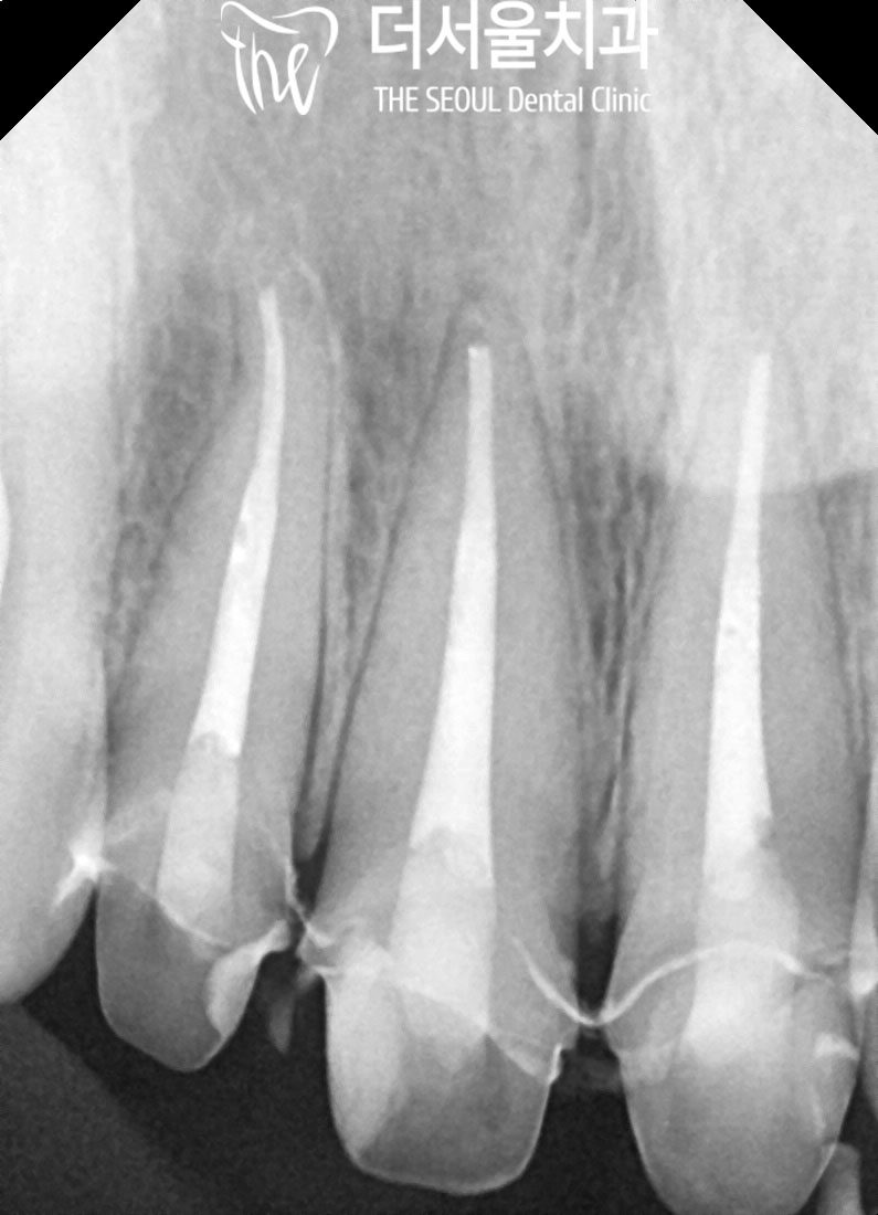
A radiograph showed the decay had extended as far as the nerve. The colour change was a consequence of that, not a separate cosmetic problem beside it. The neighbouring teeth and surrounding tissue were assessed too, by inspection, radiograph and CT: the discoloured tooth is the one the patient can see, and whether it is the only one involved is a question imaging answers rather than the mirror.
Where decay has reached the pulp, a filling is not an option that was declined — it is not available. Filling material laid over inflamed or dying pulp seals the problem inside the tooth.

Where is the hole made in a front tooth?
In the back of it.
After local anaesthetic, the decayed tissue was removed and the pulp chamber opened. On a front tooth that opening — the access cavity — is cut through the palatal surface facing the tongue, not through the surface that shows when you smile. The instruments that clean the canal need a straight path down the root, and the back of the tooth provides it while leaving the visible face untouched.
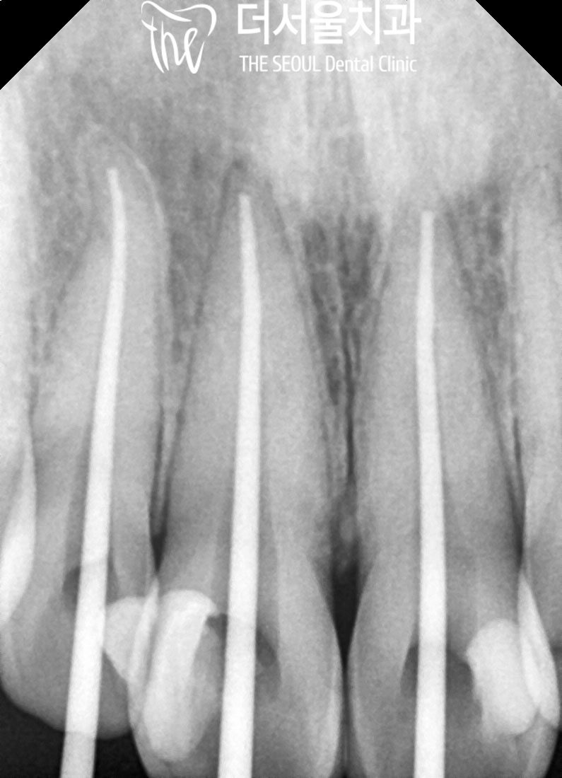
Through that opening the canal contents were removed, the canal washed and disinfected so no inflamed tissue remained, then shaped before being filled. The shaping is not tidiness: an unshaped canal cannot be filled along its full length, and space left unfilled is space bacteria can occupy again.
What did the crown involve?
A root-treated front tooth is not left as it stands, for three reasons that stack: tissue lost from inside where the pulp was, tissue lost from outside where the decay was removed, and a shade the root canal cannot restore — it removes the cause of the discolouration, not the discolouration already in the tooth.
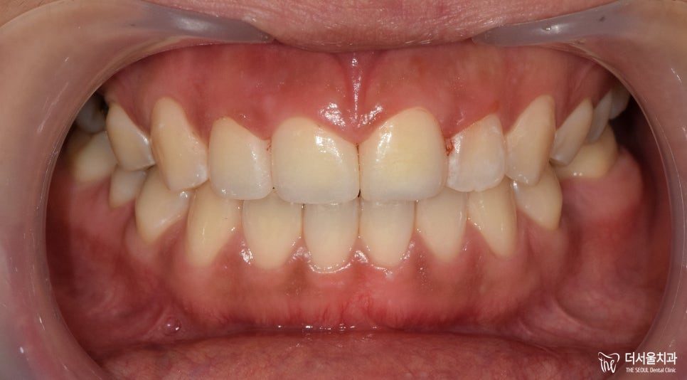
The crown of the tooth was shaped — preparation — to the thickness the material requires, and a zirconia crown was made and fitted, its shade and shape agreed before it was made rather than after it arrived.
Zirconia is one material through its whole thickness, tooth-coloured inside and out, with no metal core. Two things follow on a front tooth: no dark line can develop at the gum margin, as it can where porcelain is fused over metal, and the shade is not influenced by the colour of the tooth beneath it — which matters on a tooth that darkened in the first place. The trade-offs between materials are on our crowns and veneers page.
How long did it take, and how many visits?
This case ran from 4 October to 2 November 2024 — about four weeks from first examination to fitted crown, across separate appointments. Root canal treatment takes more than one visit: the canal is cleaned and disinfected, then filled once the tooth has settled. Preparation and the impression follow, and the crown is fitted once the laboratory has made it.
If you are visiting Korea rather than living here, four weeks with several appointments is the shape to plan around, not a slow version of a one-day treatment. Where a trip is shorter, say so at the first appointment. On cost, the root canal and the crown are billed as separate items and crown materials differ in price; our published fees for non-covered treatment are on the pricing page.
Two checks are made at fitting. The bite — how the crown meets the opposing teeth when you close and slide the jaw, since a front tooth guides that movement and a crown sitting slightly high takes force it was not shaped for. And the response to cold and heat: the treated tooth has no pulp left to respond with, so sensitivity afterwards points to a neighbouring tooth or to the crown margin.
Three reasons to have a discoloured front tooth examined
Any one of these is enough:
- one front tooth is darker than the teeth either side of it, and getting darker;
- a grey or brown shadow at the biting edge or between the front teeth, with no pain and nothing to feel;
- a front tooth filled in the past has changed shade since.
An examination with a radiograph settles which side of the line the tooth is on: still a filling, or already a root canal. Waiting does not move a tooth back across that line.
Frequently asked questions
Can a decayed front tooth be filled instead of root-treated?
Where the decay has not reached the pulp, yes. Where it has, a filling seals inflamed or dead tissue inside the tooth rather than removing it. A radiograph distinguishes the two, before the decision rather than after.
Is a tooth weaker after root canal treatment?
It has less structure than before, from the decay removed and from the access cut to the canal. That is why a crown goes over it: the crown spreads biting force across the tooth instead of concentrating it on the remaining walls.
Does the treatment hurt?
It is carried out under local anaesthetic. A tooth with an inflamed pulp can be harder to numb fully than a healthy one, so if you still feel sensation once treatment starts, say so — more anaesthetic can be given before it continues.
How long should I allow if I am travelling to Korea for this?
This case took four weeks across several appointments. A root canal and a crown are staged by nature, so the total is measured in weeks rather than days. If your stay is shorter, raise it at the first visit.