
Three teeth in the same mouth, all needing extraction and an implant, all planned on the same scan — and three quite different operations. This is why a single quoted price for “an implant” tells you very little.
What happens if a root infection is left?
Infection at the root of a tooth does not stay confined to that tooth. Left alone it affects the neighbouring teeth, and what would have been one loss becomes two or three.
It is tempting to reason that an implant can replace whatever is lost. It can — but a replacement is a replacement, and it is common to appreciate that only after the natural tooth has gone.
What did the X-rays show?
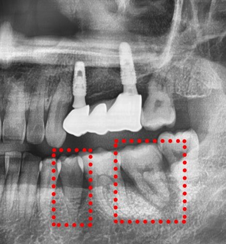
The panoramic X-ray showed dark areas at the root tips of three teeth: an upper right premolar, and two lower left molars.
Those dark areas are infection from bacterial invasion. All three had progressed far enough that treating and keeping the teeth had a poor prognosis; extracting them was what would protect the rest.
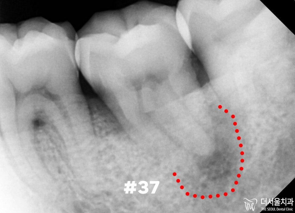
Periapical films were then taken of the individual teeth. At that magnification the lesions were far more clearly defined than on the panoramic view — which is the reason both are taken rather than one.
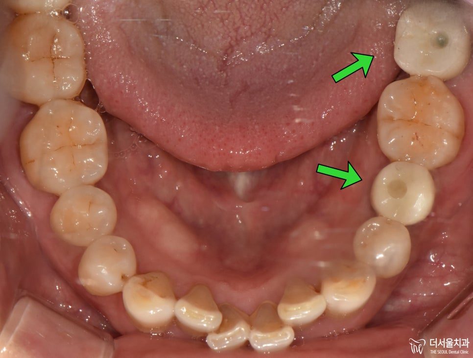
The three sites
Digital scanning and simulation gave a different assessment for each.
Upper right premolar — routine
No significant bone loss. Adequate thickness and depth. Straightforward placement.
Lower left second premolar — care required
Some bone loss, bringing the site closer to the nerve canal than ideal, but not enough to require grafting.
The specific risk here was the drill skidding during preparation. Knowing that in advance is what allows it to be prevented; discovering it during surgery is a different matter.
Lower left second molar — difficult
The infection was extensive and so was the bone loss. Very little bone remained, and what was left did not give enough vertical height to keep clear of the nerve canal.
This site needed grafting first to build a foundation before a fixture could be expected to hold.
What does a surgical guide do?
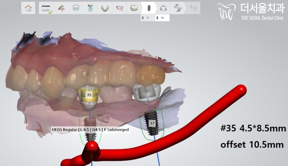
From the simulation, a surgical guide was produced to transfer the planned angle and position of each fixture into the mouth. The guide also allows minimal incisions and shortens the time each site is open.
How the sites were sequenced
The upper premolar was completed first — healthy bone, no complications.
The lower molars were given longer to consolidate. The heavily grafted site was submerged: covered over by the gum and left buried while the bone integrated, rather than left exposed through the tissue. Where bone loss is severe, keeping the site sealed protects it during the period when it is most vulnerable.
At three months, integration was confirmed, impressions were taken and the final restorations were made.
How long did the whole treatment take?
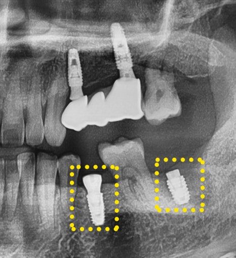
Abutments and crowns were fitted, and a final panoramic X-ray confirmed the result across all three sites.
The pain the patient had been chewing through was gone, because its cause — the root infection — had been removed with the teeth.
Treatment ran from 7 December 2023 to 18 April 2024, about four and a half months.
How do you look after an implant afterwards?
That placing the implants well is the smaller half of the job. What was covered at the final appointment was how to clean around them, how to brush, and when to come back.
An implant does not decay, and the bone around it is as vulnerable to plaque as it was around the tooth it replaced. Regular professional review matters, and so does what happens in between.
Frequently asked questions
Why do implants at different sites cost different amounts?
Because they are different operations. One site may need nothing beyond placement; another may need bone grafting, a sinus lift, or submerged healing. That is settled by the scan, which is why a figure quoted before imaging is not a real estimate.
What does it mean for an implant to be “submerged”?
The fixture is covered over by the gum and left buried while it integrates, rather than left projecting through the tissue. It is used where bone is poor or heavily grafted, to protect the site during the period when it is most vulnerable. A second small procedure uncovers it later.
Why take both a panoramic and a periapical X-ray?
The panoramic gives the overview across the whole mouth; the periapical shows one tooth at much higher detail. Lesions that are faint on the panoramic are obvious on the periapical, and the plan depends on knowing their true extent.
Should an infected tooth be extracted or treated?
It depends on how far the infection has gone. Where it is limited, root canal treatment often saves the tooth. Where the lesion is extensive and the bone around it is going, extracting protects the neighbouring teeth — which is the wider loss being weighed against the one tooth.
Can several implants be done at once?
They can be planned together and often placed in the same period, but they do not necessarily finish together. Here the straightforward site was completed first while the two lower sites were given longer to heal.
Read more: bone grafting and sinus lift · digital implants · root canal treatment