
Two words come up whenever implants are mentioned: expensive, and painful. Both have a reason behind them — conventional placement means opening the gum to reach the bone underneath. A flapless, digitally guided approach changes that part of the equation, but it is not available to everyone. Here is what decides it.
What does “flapless” actually mean?
In conventional implant surgery, part of the gum is incised and lifted so the fixture can be placed into the alveolar bone beneath. Some discomfort follows as a matter of course.
The approach described here places the fixture immediately into the socket left by an extracted tooth, so no unnecessary incision is made. Less cutting means less bleeding and, for most patients, a shorter recovery. It is not a different implant — it is a different way of reaching the same place.
How is the digital guide made?
- CT scan — the oral structure and the state of the jawbone are analysed in detail.
- Intraoral scan — the exact shape of teeth and gum is captured.
- Simulated surgery — the placement position and angle are worked out on computer before anyone touches the patient.
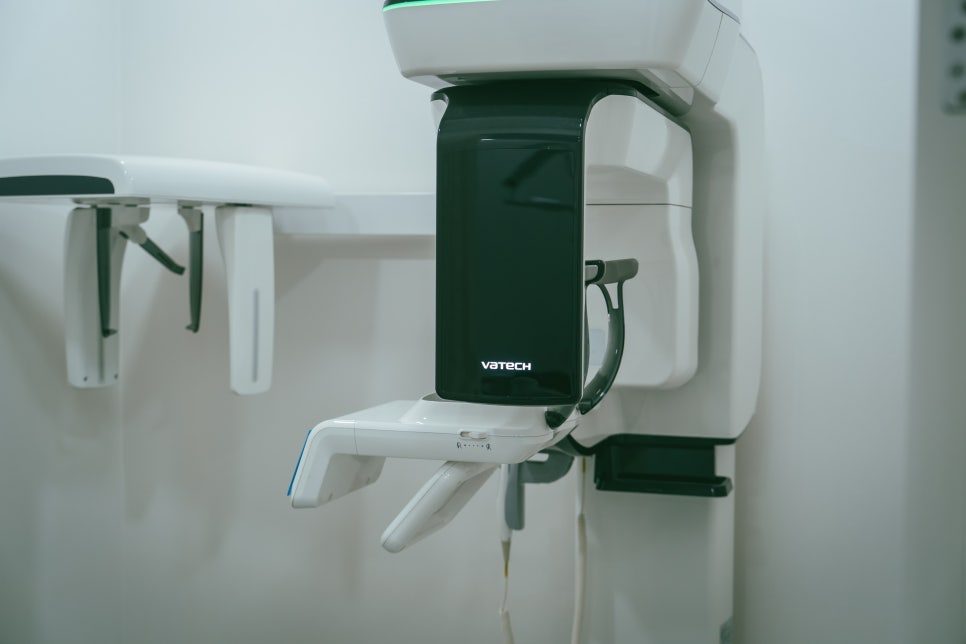
- 3D-printed surgical guide — the plan is transferred into the mouth, so the actual placement follows what was simulated.
Who can have a guided implant?
Two conditions have to be met:
- The jawbone has to be sufficient in both quantity and quality.
- The gum has to be free of inflammation or disease.
Minimising the incision requires reasonably healthy gum and bone to begin with. That said, insufficient bone does not automatically rule it out — adding a bone graft makes it possible in some cases, which is exactly what happened here.
We particularly consider this approach for patients who:
- have limited time and want treatment and recovery to move quickly
- are sensitive to pain and find conventional surgery daunting
- need visible areas treated with as little trace as possible
- are older, where reducing physical strain matters
Even when all of these apply, the final plan still follows diagnosis. None of it is decided in advance of an examination.
The case: upper molar in a patient in their late eighties
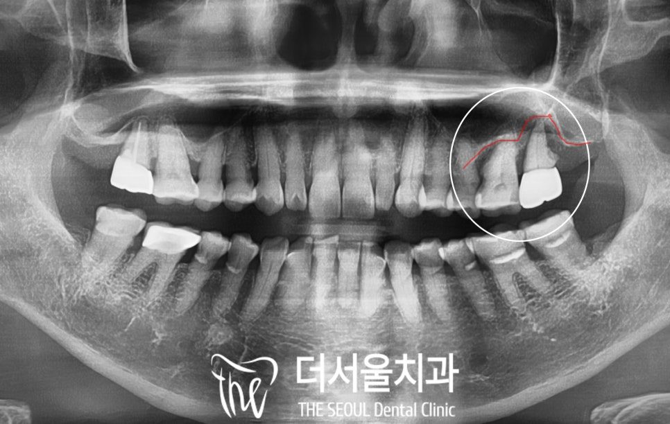
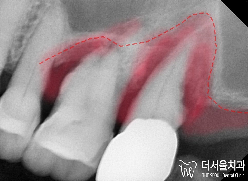
This patient fell into the fourth group above. The upper left molar area looked unremarkable to the eye, but X-rays showed significant alveolar bone recession around the root. A periapical view confirmed that the periodontal ligament holding the root and the surrounding tissue had broken down, and mobility was considerable.
There were no systemic conditions and no medications to work around. The mobile molar was extracted and a fixture placed into the site with a minimal incision.
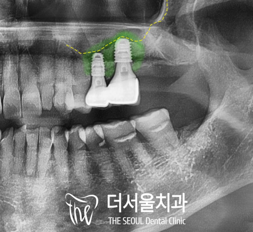
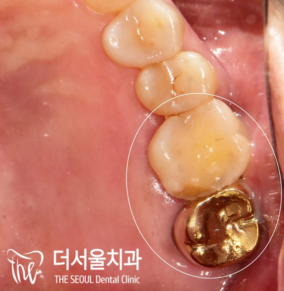
Bone loss was severe enough to make this a demanding case. A sinus lift raised the maxillary sinus membrane, and sufficient bone grafting was placed in the space created to reinforce the deficient area. On the follow-up X-ray the grafted material is visible as a radiopaque area. Several months later, once osseointegration had progressed, the final prosthetic was fitted.
Timeline
Treatment ran from February to late June 2024 — a little over four months from placement to final prosthetic. The waiting time in the middle is bone healing, and it cannot be shortened by scheduling. Patients travelling from overseas should expect this stage to need either a second trip or an extended stay, and should raise their travel dates at the first consultation so the stages can be planned around them.
Frequently asked questions
Is a flapless implant less painful than the usual method?
Avoiding an incision generally reduces bleeding and post-operative discomfort, and most patients recover faster. How much difference you experience depends on the site, the amount of bone work needed and your own pain sensitivity, so it is not a guarantee for every case.
Can I have an implant if I do not have enough jawbone?
Often yes. Bone grafting, and in the upper jaw a sinus lift, can rebuild the site so a fixture can be placed. It adds healing time to the schedule, but insufficient bone by itself is not a reason to be turned away.
Is there an age limit for dental implants?
Age alone is not the deciding factor. What matters is bone condition, gum health, systemic conditions and any medication you take. The patient in this case was in their late eighties with no systemic conditions, and treatment proceeded normally.
What is a sinus lift, and why would I need one?
The maxillary sinus is an air cavity above the upper molars. When bone height there is too low for an implant, the sinus membrane is lifted to create space, and bone graft material is placed in it. Lifting the sinus without grafting achieves nothing on its own — the graft is what gives the fixture something to hold onto.
Read more: digital guided implants · bone grafting and sinus lift · dental implants overview