
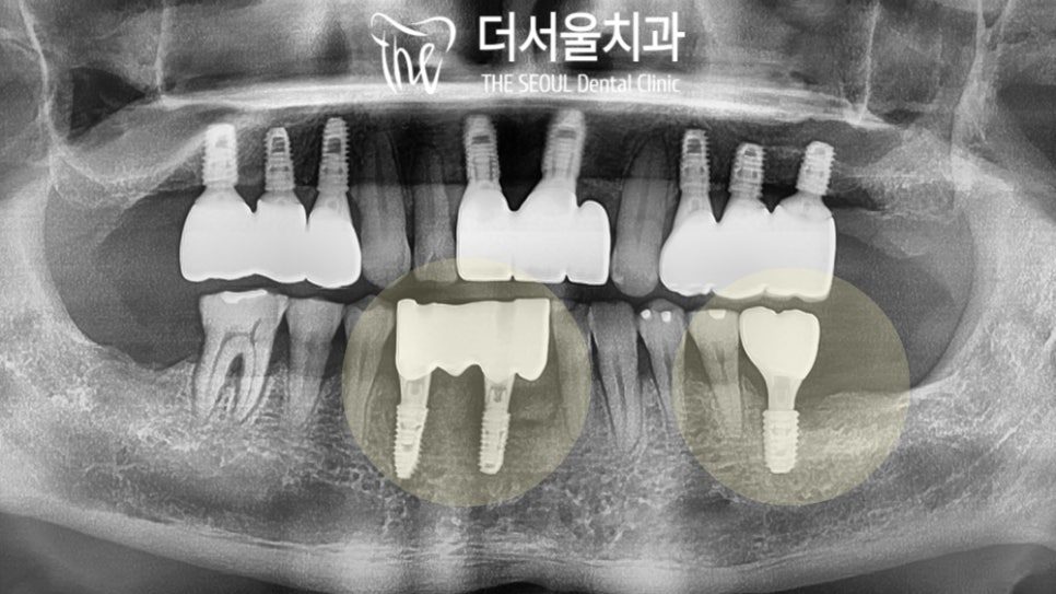
The lower front teeth give an implant less room to work with than anywhere else in the mouth. This case had a front tooth already destabilised by infection at the root end, and a missing molar on the same jaw — two very different placement problems in one treatment plan.
What the patient presented with
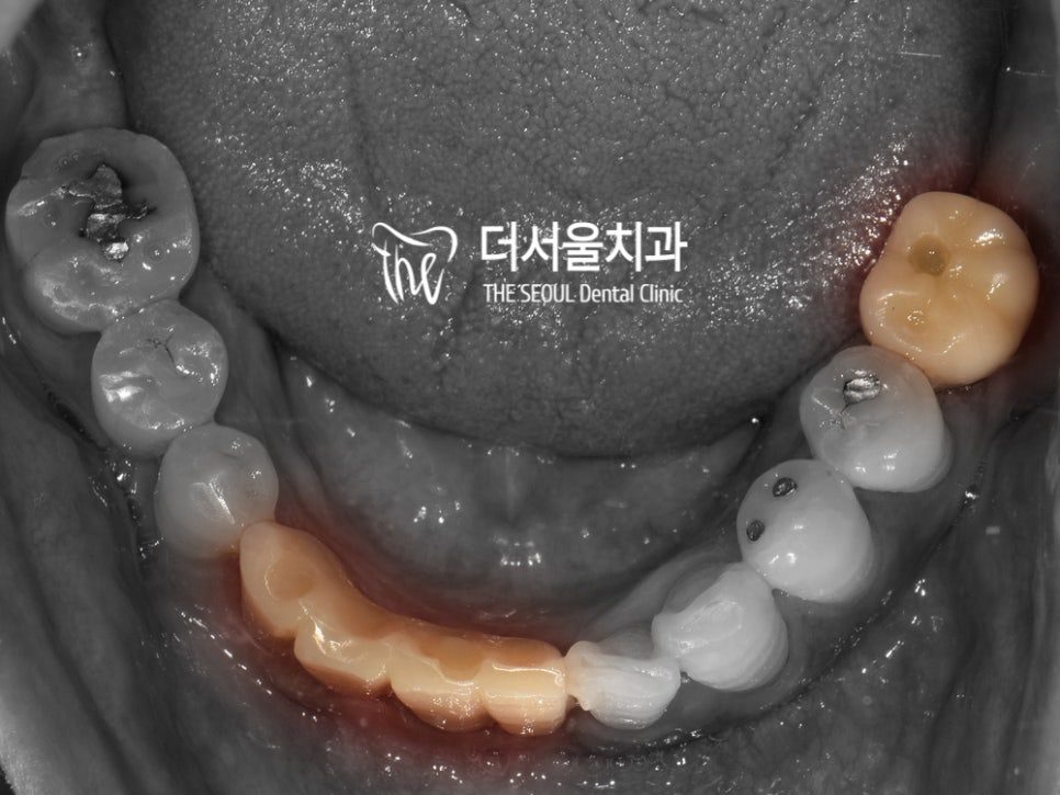
The patient came in with pain and mobility in a lower front tooth, and a molar that had been lost some time earlier. Intraoral photographs showed a single front tooth holding on precariously.
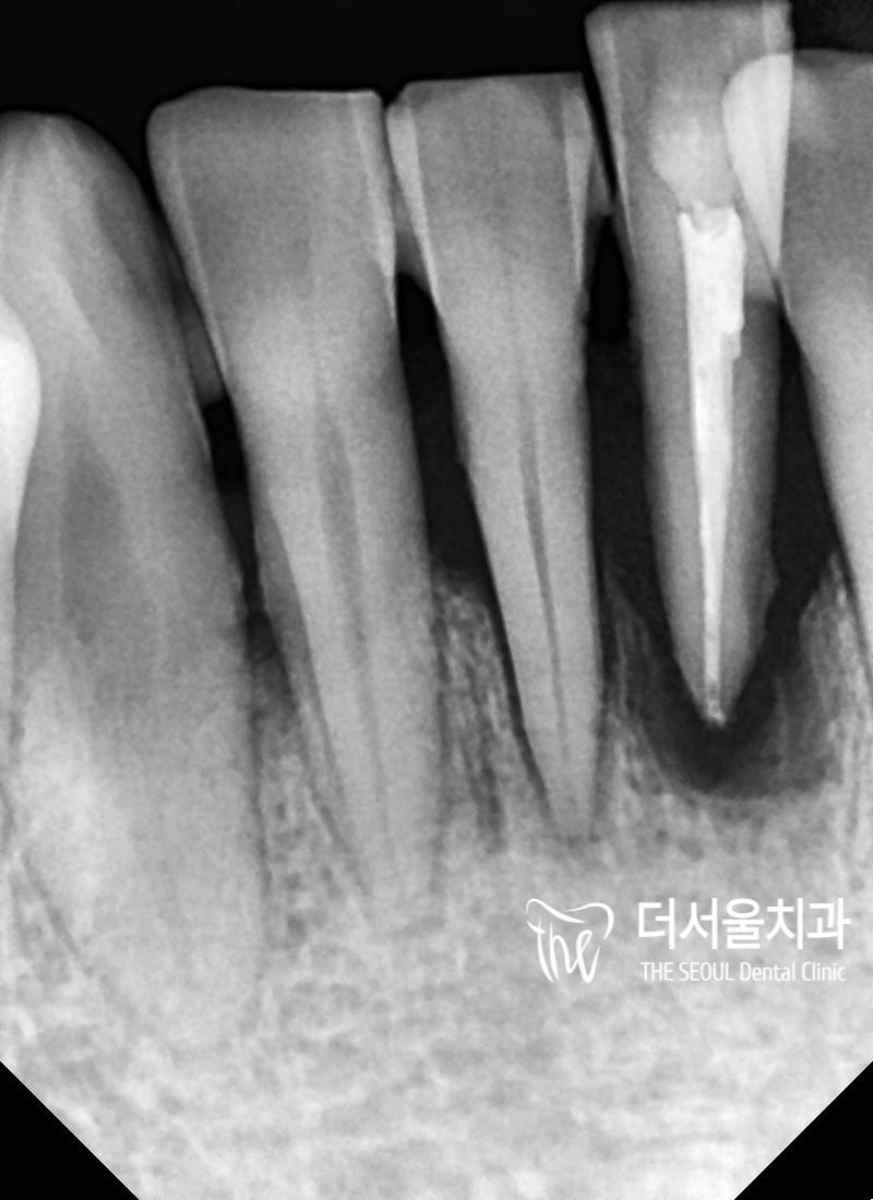
A periapical X-ray explained why. Extensive inflammation had developed at the root end and had progressed into loss of the surrounding alveolar bone. Once that bone is gone, the tooth has nothing holding it, and no treatment directed at the tooth itself restores the support.
The plan was therefore to place implants in both sites — the failing front tooth and the missing molar — to restore chewing function and appearance.
Why is the lower front the tightest site in the mouth?
Lower incisors are narrow teeth set close together, and the ridge of bone beneath them is thin from front to back. That has two consequences:
- Placement. There is very little margin between the fixture and the roots of the neighbouring teeth. The angle matters as much as the position, because a fixture that is correctly placed at the crest can still be heading towards a neighbouring root deeper down.
- The restoration. The crown has to be narrow enough to match its neighbours while still being strong enough to function, and it has to emerge from the gum at a natural profile. There is no room to compensate for an error by shaping the crown differently.
How is a narrow-space implant planned?
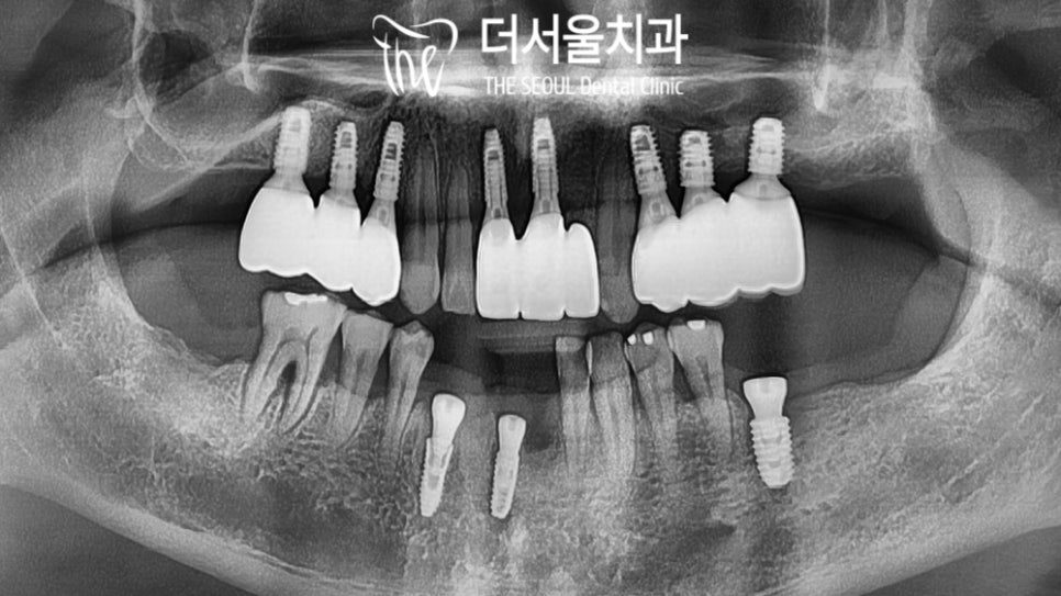
The placement position, direction and angle were determined by computer analysis of the scan and CT data before surgery, and the fixtures were placed to that plan.
Planning in advance is what makes a site like this workable. Where the tolerance is a millimetre or two, the decisions are better made on screen, where they can be checked and revised, than in the mouth during surgery.
The restorations
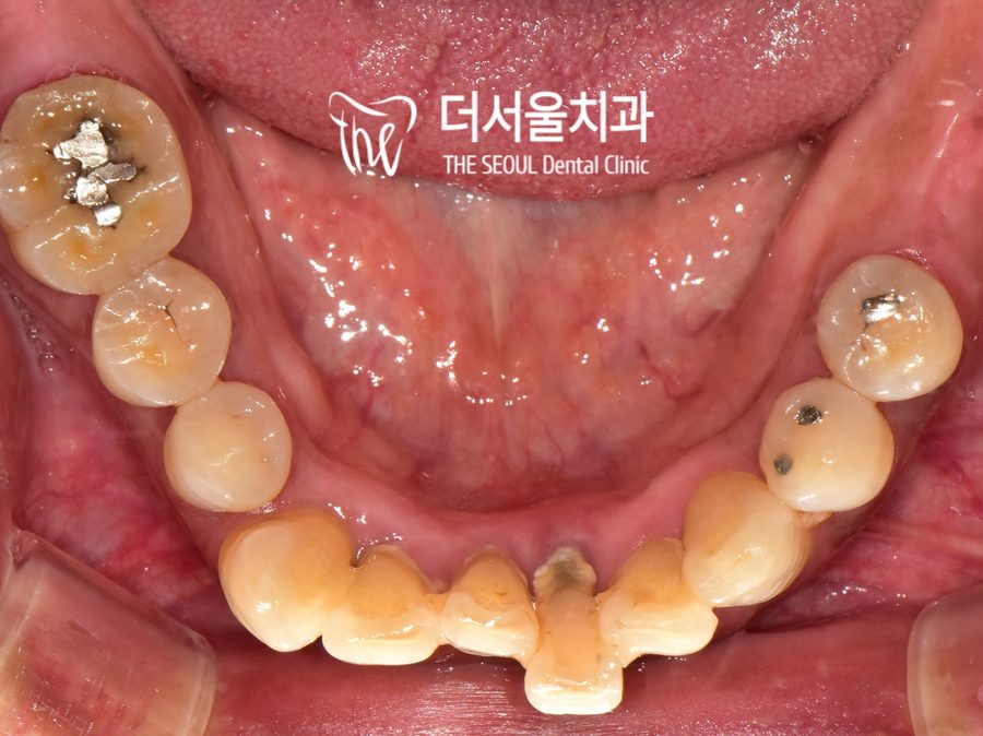
Once osseointegration was confirmed — the fixtures having bonded into the bone — the final crowns were fitted. Both the front tooth and the molar were restored in zirconia, shaped and shaded to resemble the natural teeth around them.
A final bite check was carried out before completion, so that chewing forces are distributed as intended rather than concentrating on the new restorations.
At a later review appointment several months on, both restorations were functioning normally.
Timeline
Treatment ran from late April to mid-September 2020 — about four and a half months. As with any implant case, the bulk of that is the healing period between placement and the final crown, and it is set by biology rather than by the appointment book.
How should you compare implant prices?
It is reasonable to ask what an implant costs and to compare. What is worth understanding is what the figure covers, because that is where quoted prices differ most: the fixture system used, whether the abutment is stock or custom-made, the crown material, whether bone grafting is included if it turns out to be needed, and what follow-up care is part of the fee.
Our own fees for non-covered treatment are published in English, and you receive an itemised written estimate after examination and before anything begins. We would rather you compared like with like than compared a headline number against a full treatment plan.
Frequently asked questions
Can a loose front tooth be saved instead of replaced?
It depends on why it is loose. If the cause is inflammation that has already destroyed the surrounding bone, the support is gone and treating the tooth does not bring it back. An X-ray showing the extent of bone loss is what settles the question.
Is there enough room for an implant in the lower front teeth?
Often yes, but the margin is small and it has to be measured rather than assumed. A CT scan shows the width of bone available and the position of the neighbouring roots, which is what determines whether a fixture fits and at what angle.
Will a front tooth implant look different from my other teeth?
It should not. The crown is made to match the shape and shade of the teeth beside it, and how it emerges from the gum is planned as part of the fixture position. Front teeth are the least forgiving site in the mouth, which is exactly why the position is planned digitally first.
Why do implant prices vary so much between clinics?
Mostly because the quotes cover different things — fixture system, custom versus stock abutment, crown material, bone grafting, and follow-up. Ask what is included rather than comparing headline figures, and ask for the estimate in writing.
What happens if a clinic that placed my implants has closed?
Another clinic can usually take over care, but it helps enormously to know which fixture system was used. Bring any records or receipts you have; where the system cannot be identified from records, it can often be determined from an X-ray.
Read more: digital guided implants · dental implants overview · what treatment costs