
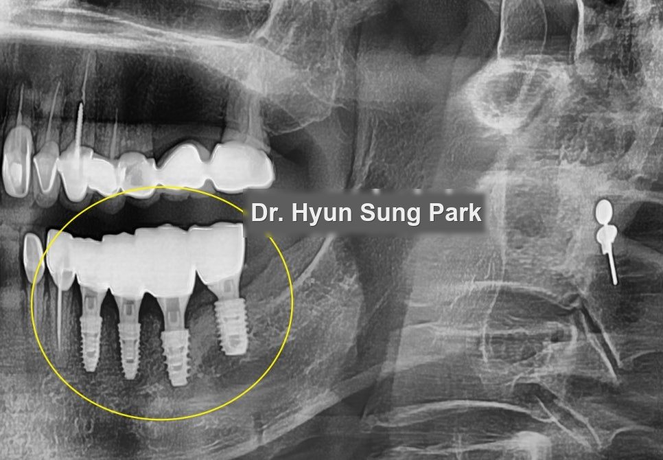
An implant that has become infected presents a fork: treat the infection and keep the fixture, or remove it and start again. Both are done. They do not have the same odds, and the deciding factor is visible on an X-ray.
How much bone had been lost?
Three implants in place. At a glance their angulation looked reasonable.
The bone around them was the problem. The bone level had dropped well outside the normal range, and on a higher-resolution image the extent was unambiguous: roughly the upper half of each fixture was no longer held in bone.
That is the number that decides the case.
Should the implant be cleaned or replaced?
1. Antibiotics and cleaning the fixture
Control the infection with medication and clean the exposed fixture surface, leaving it in place.
For: no removal, no second surgery.
Against: infection around an implant, once established, rarely stops of its own accord. The realistic expectation is that bone loss continues, and continuing bone loss around a fixture that has already lost half its support does not end well.
2. Remove and replace
Against: it means surgery again, which nobody wants.
For: it removes the problem that option one leaves in place.
Which route to take is the patient’s decision. What should inform it is how far the bone loss has already gone — and at half the fixture length exposed, cleaning was not an appropriate choice here regardless of preference. This case took the second route.
Removing an integrated fixture is not unscrewing it
This is the part patients are surprised by.
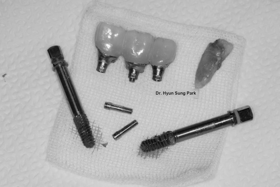
An implant that has integrated cannot be reversed out like a screw. It is fused to the bone along its surface, so it has to be removed together with the bone it is fused to — using a trephine bur, a hollow cylindrical cutter that cores out the fixture and the immediately surrounding bone as one piece.
Which means removal necessarily creates a defect. Guided bone regeneration (GBR) follows as part of the same procedure, not as an optional extra: the site has to be rebuilt before anything can be placed into it again.
The soft tissue was incised first to expose the fixtures, which confirmed what the X-ray had shown. A premolar in the same area was also in poor condition and was extracted during the same procedure.
Why is planning harder the second time?
Re-treatment raises the stakes on planning, because the site is no longer the one that was originally worked with.
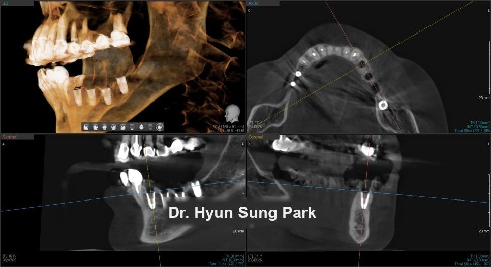
The remaining bone and soft tissue were assessed and the new fixture positions determined from that, with the surgery simulated on screen before it was carried out. Placing three fixtures into a site that has just been grafted leaves very little room for adjustment during surgery.
How was the new placement confirmed?
Three fixtures were placed in line, following the simulation.
A CT scan was taken immediately afterwards to confirm they had gone in where they were planned to go. That check matters more in a revision case than in a first placement, because the bone being worked into is grafted rather than native.
Three to four months of integration followed.
Finishing
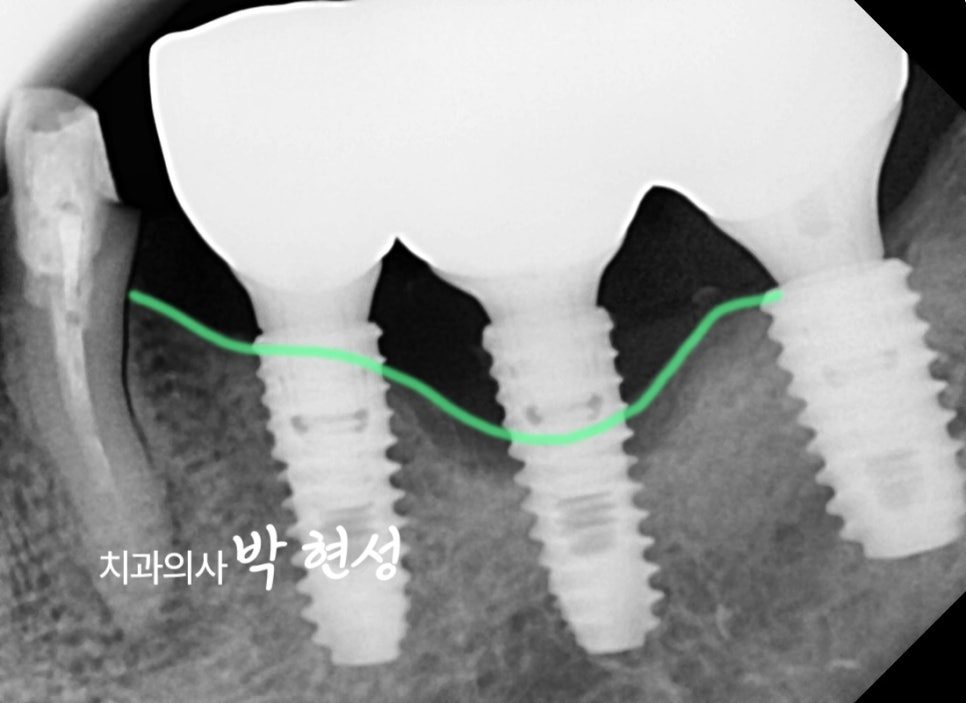
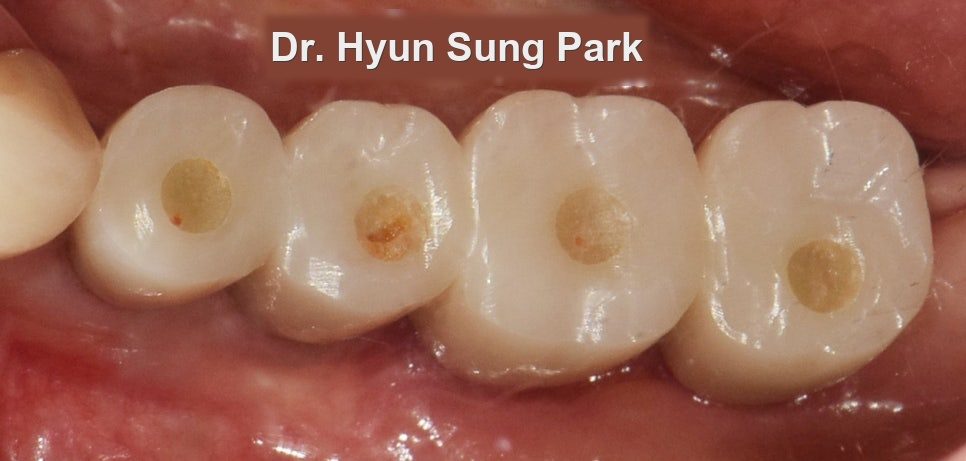
After the integration period the site was restored with a bridge across the three fixtures.
How do I stop this happening again?
Peri-implant infection is a maintenance failure more often than a surgical one. A revision case should be reviewed roughly every three months, not annually.
The reason is straightforward: bone loss around an implant is painless until it is advanced, exactly as it was the first time. Nothing about having had one implant fail makes the next one announce itself any earlier.
Frequently asked questions
Can an infected implant be saved?
Sometimes, where the infection is caught early and bone loss is limited — antibiotics plus cleaning the fixture surface. Once a substantial proportion of the fixture is no longer held in bone, that approach is unlikely to hold, and removal with grafting and replacement is the more reliable route.
How is a failed implant removed?
Not by unscrewing it. An integrated fixture is fused to the bone along its surface, so it is cored out with a trephine bur together with the bone immediately around it. That is why bone grafting is part of the removal rather than a separate decision.
How long before a new implant can go in?
In this case the fixtures were placed at the same procedure, with grafting, followed by three to four months of integration before the bridge. Where the defect is larger, grafting and placement are sometimes separated, which adds several months.
Why did the infection happen in the first place?
Most commonly, plaque accumulating around the implant. An implant does not decay, but the tissue and bone around it respond to bacteria much as they do around a natural tooth — and unlike a tooth, it gives you no early warning.
How often should I have implants checked?
Every six months for a straightforward case, and about every three months after a revision. The checks exist to find bone loss while it is still measurable in millimetres rather than in halves of a fixture.
Read more: implant revision · bone grafting · dental implants