

Most people who have a wisdom tooth removed in their fifties were told about it in their twenties. It never hurt, so no particular week was ever the week to deal with it. What worries them when they finally decide — often by then in a country not their own — is whether they have left it too long. The tooth has not changed in thirty years. The bone around it has.
Why does a wisdom tooth get harder to remove with age?
The difficulty of an extraction is mostly a property of the bone, not of the tooth.
In the late teens and early twenties, jaw bone is less mineralised and more elastic. It gives slightly as the tooth is eased against it, so the tooth can often be delivered with modest force and little bone removal. Roots at that age are frequently still forming, which leaves less root surface gripping the socket.
With each decade the bone becomes denser and stiffer, and has to be worked around rather than pressed against. By then the roots are fully formed, and they can be blunt, curved or splayed — nothing useful to lever against.
That has three practical consequences:
- The appointment takes longer.
- The tooth is more likely to need sectioning and a gum flap rather than being eased out whole.
- There is more swelling afterwards, and healing is slower than it would have been at twenty.
None of this makes removal in later life unwise. It makes it a planned procedure rather than a quick one.
The case: a lower wisdom tooth lying on its side
The patient was a man in his fifties, with no pain and no swelling. He had known for years that he had an impacted wisdom tooth and came to have it dealt with rather than because anything had gone wrong.
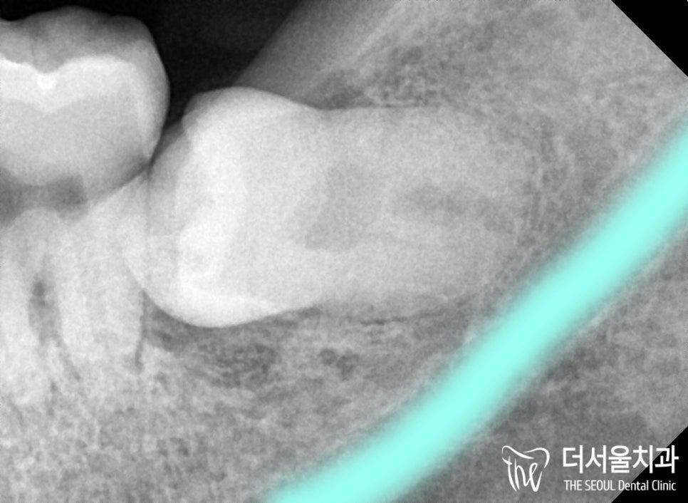
The panoramic film showed tooth 38 — the lower left wisdom tooth — lying completely horizontal beneath the gum, its crown pointing forwards into the molar in front. None of it had erupted, so a gum incision was unavoidable.
The procedure was carried out under local anaesthetic. A horizontally impacted lower tooth is more uncomfortable than a straightforward extraction, and that is worth saying plainly rather than calling the appointment painless.
How was it decided that a CT scan was not needed?
For a lower wisdom tooth, one measurement decides the plan: the distance between the roots and the inferior alveolar nerve canal, which carries sensation to the lower lip and chin. It is a sensory nerve, so injury there causes altered sensation rather than weakness.
That region of the panoramic image was examined under magnification, and the roots sat clear of the canal, so the removal could be planned without further imaging. Where roots and canal appear to overlap, a flat film cannot tell whether they truly touch or merely pass in front of one another, and a 3D CT scan is taken first.
Age is part of why that check matters: dense bone has to be worked against more firmly, and firm work near a structure whose position is unknown is exactly what needs designing out.
Why the tooth had to be cut into pieces
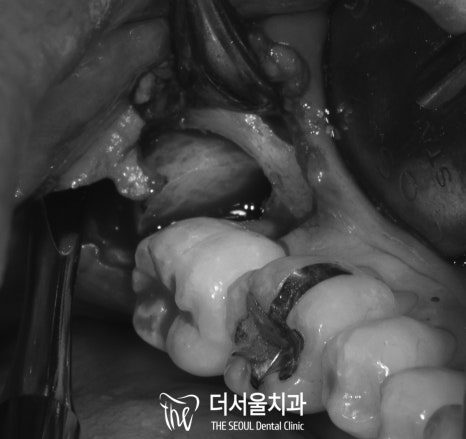
Once the gum was laid back, the tooth was sitting deep with no path out as a single unit. In more elastic bone a tooth in that position can sometimes be rotated free; here it could not.
At that point there are two options: remove bone to create a path, or reduce the tooth to fit the path that exists. The second is preferred, because bone removed to make room is bone that then has to heal. So the crown was separated first and lifted out, and the remaining root portion followed through the space the crown had occupied. The roots were blunt rather than tapered, giving no easy purchase, so each piece came out on its own.
Sectioning is not a sign that an extraction has gone wrong — it is what allows the bone to be left alone when the tooth will not come out whole. The X-ray afterwards confirmed an empty socket with no root fragment left behind.

What the recovery involved
The gum was closed with sutures, and a review appointment followed within the week to remove them and inspect the socket. It healed without inflammation.
The part patients control is the first few days. Where the instructions are not followed — the clot disturbed by rinsing or spitting, smoking resumed early — the socket can become inflamed.
For planning purposes that is two appointments: the extraction, and a review a few days to a week later. If you are visiting Korea, allow at least a week between arrival and departure for a case like this, and give your dates at the consultation so both fall inside them.
Is it too late if you are already in your fifties?
No. Difficulty rises with age; it does not become impossibility. What rises with it is the value of the planning done first.
The argument for doing it now rather than in five years is the one that applied at twenty, with less margin. The bone will be denser at sixty than at fifty-five, and healing capacity does not improve with waiting.
Three situations make an assessment worthwhile:
- You know from an old X-ray that you have an impacted wisdom tooth, and nobody has looked at it since.
- Food packs in behind your last molar, or the gum there swells from time to time and then settles.
- You have already been advised to have one removed and have been postponing it for years.
None of the three is an emergency. All three get harder to deal with the longer they are left.
Frequently asked questions
My wisdom tooth has never hurt. Is that a reason to leave it?
Absence of pain says nothing about position. A horizontally impacted tooth is covered by gum and bone, so there is often nothing to feel until it decays the molar in front or the gum around it becomes inflamed. The decision is made from imaging, not from symptoms.
Is removal riskier in your fifties than in your twenties?
The anatomy is the same and the nerve is located the same way. What differs is that denser bone makes the extraction longer and more likely to need sectioning, with more swelling and slower healing. Age-related medical factors, such as blood-thinning medication, are also reviewed beforehand.
What does it mean for a wisdom tooth to be “impacted”?
That it has not come through into the mouth, because there is no room for it or because it is angled the wrong way. A horizontal impaction, as here, means the tooth is lying on its side under the gum with its crown pointing at the molar in front.
How many visits does it take, and how far apart?
Typically two: the extraction, and a review within the following week for suture removal and a check of the socket. Here the tooth came out in a single appointment despite being fully impacted.
What happens if I keep postponing it?
The tooth stays where it is and the bone around it keeps getting denser, so the extraction becomes longer and more surgical. Meanwhile an impacted tooth can decay the molar in front of it — a tooth worth keeping — where neither you nor a toothbrush can reach.
Read more: patient cases · emergency dental care · planning dental treatment in Korea